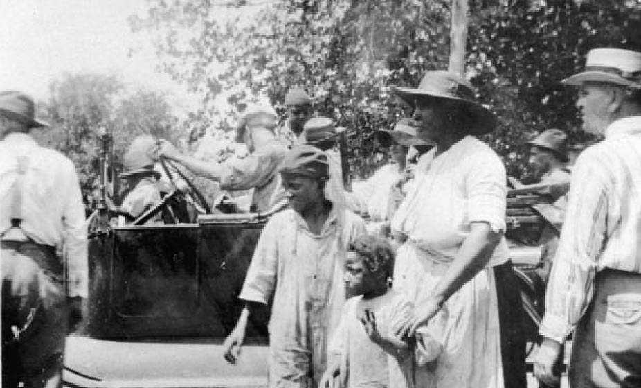
(Courtesy Department of Special Collections, McFarlin Library, University of Tulsa).
History Uncovered:
Skeletal Remains as a Vehicle to the Past
By Phoebe Stubblefield and Lesley M. Rankin-Hill
I am invisible, understand, simply because people refuse to see me.
--Ralph Ellison
Overview
During the last 20 to 30 years, several large and numerous small African American skeletal populations have been studied by physical anthropologists. Each population has contributed significantly to the reconstruction of African American lives, experiences, communities, and historical events. African Americans to a great extent are the "invisible people" in the historical record. This is a common problem whenever one studies non-elite people in the historical past, especially members of the underclass. These are the people who facilitated the lives of the wealthy and the powerful of society; they built cities, provided goods and services, and, to a great extent, were the essential elements of a growing society. However, they remain obscure in publications of their times and the history books. Elites leave significant documentation of their lives in a variety of forms and these materials have a high probability of being archived. The few sources of documentation for the poor and under classes of a society are likely to be lost.
Therefore, when African American skeletal populations are discovered or recovered they present a unique opportunity to add to the historical record and document the lives of the individuals and their community. Physical anthropological studies provide a direct method of assessment (providing evidence) when skeletal populations like the New York African Burial Ground or the Dallas Freedmen's cemetery become available.
African American skeletal populations have become available under several conditions: 1) the intentional excavation due to land redevelopment or threat of environmental damage; 2) the accidental discovery of an abandoned cemetery; 3) archaeological excavation projects for historical/anthropological research and documentation. These skeletal populations, represent a broad spectrum of African American lifestyles throughout the eighteenth, nineteenth, and twentieth centuries in the Western Hemisphere.
Biological and behavioral factors affect the human skeleton because the skeleton is a dynamic system, that undergoes growth and development throughout the individual's life span. In general, these biological and cultural factors can interfere in the normal processes of bone growth and loss, causing disease episodes and/or periods of delayed growth. These experiences can be usually indelibly recorded on the skeleton and dentition. Through observing these "historical remnants" of bones and teeth, the physical anthropologist has a means of measuring a population's health. In addition, the skeleton can record the actual cause(s) of death and/or contributory factors surrounding death.
Therefore, the potential contribution and importance of the Tulsa Race Riot victims' skeletal remains would be significant to both the documentation of the historical event and to African American history. It is imperative that these remains be located, recovered, "given a voice" through skeletal analysis, and then reinterred with dignity, as most of the African American skeletal populations have been and will be in the future.
A discussion of the basic types of analysis and information that physical anthropologists and forensic anthropologists can provide is presented below.
The Role of Forensic Anthropology in the Identification of Deceased Individuals
Forensic anthropology has had an active role in American science and medicolegal investigations since at least 1878, when Harvard anatomist Thomas Dwight published his essay on identifying human skeletal remains.1 Existing as a poorly recognized subfield of the scientific discipline called physical anthropology, forensic anthropology received little scholarly or public notice until the task of identifying and repatriating the deceased from World War II and the Korean War brought the field into prominent activity. Technical advances at this time and a steady increase in academic interest in the field led to its later organization as a section of the American Academy of Forensic Sciences in 1972. Since that time, forensic anthropology has been a recognized subfield of physical anthropology and the forensic sciences, requiring the usual academic rigors of obtaining the higher degrees in anthropology (at least a Master's degree), as well as the special training and certification of its section in the Academy.
A forensic anthropologist is a physical anthropologist who has been trained to recognize and examine human skeletal remains for indications of sex, age, height, unique characters of the individual, features which might indicate how the person died, and processes that affect the skeleton after death. Although a forensic pathologist or other medical doctor may seem a more appropriate conductor of such analyses, their education and training focuses on changes in soft tissue. The forensic anthropologist is expected to recognize bone outside of its natural context even if it is reduced to small fragments. He or she can identify all the bones of the human skeleton, determine if a bone is human or not, and understand that the shape of a bone is related to its function in the body and its owner's relationship to other animals.
Forensic anthropologists serve the public in several types of investigations. As a result they work with the other agents concerned with the disposition of human remains, such as medical examiners or coroners, local and federal law enforcement and family organizations. The most common circumstances are criminal investigations on a local or federal level, such as a local homicide or the results of terrorist activity. Other circumstances include mass disasters of natural or human cause, such as the recovery of tornado or aviation accident victims. The U.S. Army maintains a staff of forensic anthropologists at a facility based in Hawaii who are dedicated to the continued recovery and identification of Americans lost in the past armed conflicts. Frequently the public learns of the forensic anthropologists work when it involves cases of historical interest, such as the exhumation of President Zachary Taylor for an investigation of the cause of his death, or the recovery and identification of the remains of the last Czar of Russia and his household.
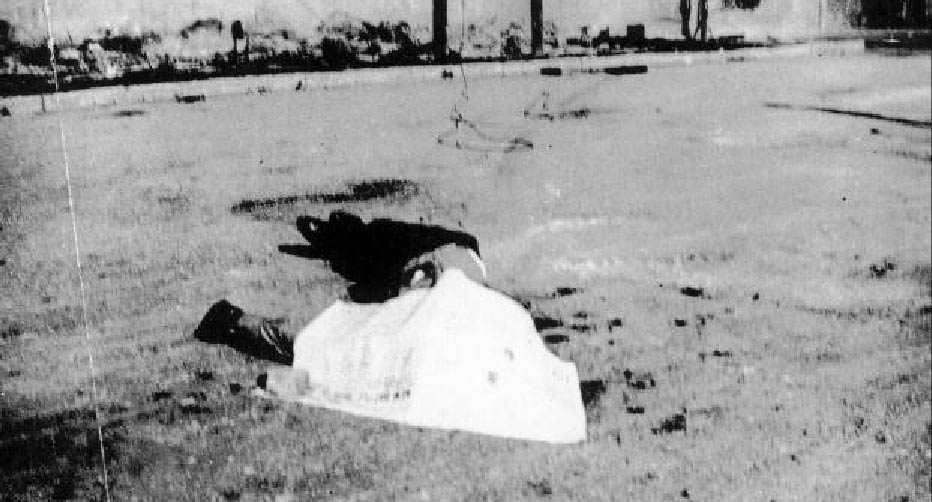
While stories have persisted for years that the bodies of riot victims were thrown into the Arkansas River, there is little evidence to support this oral tradition. Considerable oral and written evidence does exist, however, which points to African-American riot victims being buried in unmarked graves at Oaklawn Cemetery, Booker T. Washington Cemetery, and perhaps, Newblock Park (Courtesy Department of Special Collections, McFarlin Library, University of Tulsa).
The varieties of occasion that require the skills of a forensic anthropologist are sufficiently diverse that the anthropologist may enter the project at various points and utilize a wide assortment of skills. The list below is a summary of exercises that could be employed in a generic investigation. While it seems a short list, many activities take place under each section. While all of the items listed will be covered, most of the remainder of this chapter will focus on item three, laboratory analysis.
1. Scene or locality search for skeletal remains or burials
2. Recovery of remains by surface recovery or excavation
3. Laboratory analysis
4. Report production
As previously stated, forensic anthropologists are trained to discriminate between human and non-human bone. In many investigations, the anthropologists services begin and end (if no human bones are found) at this step when he or she is called to a locality or medical examiner's office and asked to make a determination. At an investigation scene the forensic anthropologist will search for and identify human bone, look for indications of burials, and conduct necessary excavations in a systematic manner using thorough documentation. In the search for burials, in addition to using visual clues, the anthropologist may employ specialized equipment and techniques, such as ground penetrating radar and infrared photography.
As part of recovery of remains, the anthropologist may map the locality in order to have a record of the position of the remains relative to a fixed landmark and any significant features of the site. This is a typical part of a criminal investigation and can be conducted in conjunction with scene investigators. Locating the site on an existing map and noting the physical address of the location may suffice, but in wooded areas or along roadsides the anthropologist may employ a Global Positioning System (GPS) unit to get the geographic coordinates of the site. If a burial is involved the site must be mapped with the location of the burial indicated (sometimes the burial is the site), while the burial itself receives a mapping grid. The grid provides a means of mapping the location of each bone or artifact found within the burial. An organized and thorough excavation may provide the information that allows the reconstruction of the events surrounding the burial of the deceased. In one instance the late Dr. William Maples successfully documented differing times of death for multiple individuals in one grave, based on the information gained from his thorough excavation.2 In addition to any physical mapping of the burial, good note taking, photography and/or videotaping during the excavation also will ensure a good record of what was found during the excavation.
Once human remains are found, they are collected in a manner that will protect the privacy of the family of the deceased, keep material remains in association, and prevent fragile material from further breakage or deterioration from exposure to air and sunlight. The remains are then maintained in a secure location while the anthropologist conducts the analysis. Good security ensures the remains and any items with them stay together and are not adulterated or altered by outside influences.
What Skeletal Remains Tell Us
In everyday living, our skeletons are frames from which we work our muscles, frames which we protect from breaking whenever possible and rely on as silent partners as we move through and manipulate the world around us. Yet human bones are not just a frame for the flesh, they also are frames for our identities. An anthropologist can get more information from a skeleton with all of its parts present, no bones broken, and little or no degradation from the environment. Even fragmentary remains will tell much about their former owner. Forensic anthropologists investigate six properties when examining skeletal remains: age, sex, ancestry, stature, unique characters of the skeleton, and indications of trauma.
Age Assessment
Unless the skeleton is sparsely represented, forensic anthropologists do not rely on only one technique to arrive at an age assessment. The best assessments are a summary conclusion based on as many parts of the skeleton as possible. This technique becomes especially important when dealing with mature individuals, because they have fewer age-specific characters than infants, children, and young adults.
Age Determination in Infants, Children, and Young Adults
The techniques for determining skeletal age in children are based on standards of skeletal and dental maturation developed for living children. Infant remains are aged by comparing the length of the long bones of the legs or arms to guidelines for the maturation of living infants. One difficulty in aging infant remains is that their bones are very fragile, do not preserve well under ground, and are rarely recovered from burials. Older children, depending on how far into development they are, can be aged by various techniques, including long bone length, degree of completed growth of the teeth, and degree of completed growth of the long bones. Age assessments using dental remains are primarily based on the degree of development of each tooth crown and root, the simultaneous presence of adult and baby teeth, and whether a tooth has erupted and if so how far. This technique is useful from infants with teeth still developing inside the jaws, to teenagers with developing wisdom teeth. The dental eruption sequence may alone be enough to obtain an age assessment, but eruption of the wisdom teeth cannot be considered an indication of adulthood because their eruption times are highly variable.
The long bones of the arms and legs each have a main shaft that develops ends that fuse as the person matures. The age that the ends develop and fuse to the main shaft occurs so regularly that age can be assessed within a couple years if enough of the skeleton is present. Limb bones stop being useful for age assessment in early adulthood. The bones in the arm, being the last to fully develop, do so at about 18 years in women and 19 years in men. As a general rule when confronted with a skeleton that looks mature on first glance, the collarbone is examined first. The collarbone is the latest fusing long bone, becoming complete by about 25 years in males and females. If the collarbone is completely united, the anthropologist uses techniques for aging adult remains.
Age Estimation in Adults
Assessing age in the adult skeleton presents a special challenge because any parts that were going to fuse as a part of maturation have done so. Most standardized techniques for age assessment in adults focus on age related changes to mature bone in portions of the post-cranial skeleton. In 1920 and again in 1989, anthropologists published standards for age changes at the fibrous joint between the pubic bones, tile pubic symphysis.3 Similarly, in 1986, anthropologists began publishing standards for the age changes to the sternal end of the fourth rib.4
Quite frequently a skeleton is too fragmentary or too poorly preserved to retain the pubic bones or the fourth rib. In such a case more marginal age estimation techniques may be used such as closure of the cranial sutures. Contrary to popular belief, cranial suture closure, as seen by the disappearance of the lines separating the bones of the cranium, is one of the most unreliable techniques for estimating age. Cranial sutures do not close in a systematic fashion in any human population. As a result, an age estimate of 30 to 50 years is not uncommon from this technique, which only signifies that the remains are adult, as was already known. Cranial sutures are used only as a last resort, such as when only a cranium is found.
In addition to using the suitable standardized techniques for the skeletal remains, the anthropologist also examines all the collected remains for general indicators of age. He or she examines the teeth, to see how worn or decayed they are in order to assess how long they were in use. Tooth wear is a population dependent character because some populations use their teeth as tools, get more dental care, or eat more grit than others. The joint surfaces and vertebrae also are examined for signs of arthritic development. In general, an older body will show more signs of lost cartilage and have more extensive bony growth on the margins of the joint. Vertebrae in particular begin developing bony growths called osteophytes as a person enters his or her 30s. The osteophytes increase in size and number as a person grows older. Another indicator of greater maturity is the presence of ossified soft tissue, such as the thyroid and cricoid cartilage of the throat, the cartilage joining the ribs to the sternum, and sclerotic portions of the descending aorta. As stated earlier, every suitable method, beginning with the most reliable, should be used for an age assessment, but forensic anthropologists are especially careful while using qualitative clues. An overused and overworked body will have arthritic development and ossified soft tissue at a younger age than otherwise expected.
Sex Assessment
It is extremely difficult to estimate sex for pre-pubertal remains because the characters of the skeleton that indicate sex do not appear until after puberty. A few techniques have been proposed for estimating sex in infants, but the reliability of these techniques is questionable. Hunt and Gleiser (1955) developed a technique for children age two to eight, based on a combination of dental and skeletal development of the hand and wrist. This technique works better than 50 percent of the time, but does require a fairly intact skeleton.
For adult remains, estimating sex can be one of the simpler parts of a forensic analysis if certain parts of the skeleton are present. Given a choice, a forensic anthropologist would always prefer to have an intact pelvis, with the second choice being an intact skull. For either part two approaches are used to estimate sex, a morphological assessment and/or a metric assessment. The morphology or shape of the pelvis differs between males and females. This difference can be recorded by noting the presence of features associated with a particular sex, or by measuring the pelvis and using statistical analysis to estimate sex.
Forensic anthropologists understand that the sex differences in the human pelvis are related to differences in function and are trained to recognize the physical differences associated with function. The female pelvis differs from the male in being designed to pass a large brained infant through a narrow space. The pelvis is made of three bones, the two innominates plus the sacrum. The innominates, themselves are composed of three bones that fuse at about age 13 in girls and 15 in boys, the pubis, ischium, and ilium. As a means of orientation, consider that when you sit down on a firm surface the bone that makes contact is the ischium, the bony hip you rest your hand on is the ilium, and the part that may unfortunately connect with the bar on a mens bike is the pubis. The female pelvis differs visibly from the male by having, among other features, a rectangular shape to the body of the pubis, a wide sciatic notch between ilium and ischium, and a pronounced angle beneath the body of the pubis.
In contrast to the pelvis, sex differences in the skull make males exceptional. Larger size plays a part here rather than a different shape, because while skulls serve the same function no matter the sex, men tend to be larger and or more robust than are women. Greater robusticity means that in the male skull projections protrude farther, and ridges are rougher and sharper. In the skull, the male brow tends to project farther than in females, and the mass of bone behind the ear, the mastoid process, tends to be larger. Size and ruggedness also will distinguish male long bones and vertebrae.
Forensic anthropologists do not rely solely on morphology to estimate sex because there are several circumstances when this technique is insufficient. Skeletal remains are frequently fragmentary. Also, differences in size and shape occur as central tendencies surrounded by variation. Therefore, we can say that the female pelvis has certain features, but we do not expect every female pelvis to have all those features in the same degree. In addition, human populations differ in the degree to which males are more robust than females. Consider the contrast of the American quarterback with his cheerleader girlfriend juxtaposed to the Eastern European bride. The alternative to, or support for a morphological assessment is to compare measurements of the pelvis, skull, or other parts of the skeleton to statistical samples generated for particular populations. The equations of Giles and Elliot are frequently used to determine sex for skulls from Americans of European and African descent.5 Statistical procedures are very important in the next two points of a forensic identification, ancestry and stature.
Determining Ancestry
The skull is the best source of information for estimating ancestry from the human skeleton. Just as with the pelvis in sex assessment, morphological and metric analysis of the skull can show the geographic population to which an individual belonged. A geographic population is the large collection of people such as Europeans, Africans, and Asians that is usually called a "race." Here the term race is avoided because the skull only indicates genetic ancestry, not the social connotations of race. Social issues of race such as "passing," or "one-drop rule," are rarely represented by the shape of the skull. In the same way that someone resembles his or her other relatives, that resemblance carries down to the bone and can be approximated with measurements and careful observation. When assessing ancestry we frequently state it in terms of descent. Typically in the United States we encounter individuals of European, African, Asian (which includes Native Americans), or mixed descent. This does not mean that the individual in question recently immigrated to the United States; rather, it means that the person's ancestry is derived from that population.
Forensic anthropologists determine ancestry by examining the morphology of the skull and by taking measurements at several points on the skull. In a morphological exam the anthropologist looks for particular sets of anatomical features that are found with greater frequency in certain populations. Closely related people will share more cranial features with each other than with their more distant relations on the next continent. On the other hand, since large populations are not made up of clones, the anthropologist cannot expect everyone in a particular population to have the same features in the same degree or combinations. Also, since all humans are related, the anthropologist cannot expect any cranial feature to necessarily be exclusive to a particular population. Therefore, an assessment of ancestry is based on a suite of characters that tend to appear or are found in similar degree in particular populations. For example, the anthropologist might look for a short, high cranium combined with a narrow nasal aperture as part of an indication of European ancestry, but he or she would not require a short, high cranium because some Europeans have long craniums. Nor would we look only for the ratio of skull length to height because different populations can have the same ratio. See the table below for a list of some of the characters used for determining ancestry.
In addition to the morphological assessment, the forensic anthropologist can conduct a metric analysis of the skull. A metric analysis requires that a skull be measured across several points, and those measurements compared to a statistical sample of individuals of known ancestry. In the United States many forensic anthropologists rely on another set of equations designed by Giles and Elliot that distinguish between people of European, African, and Native American descent.6 Anthropologists at the University of Tennessee also have produced a statistical package called FORDISC that serves a combined function of ancestry and stature estimation. Metric analysis is often the preferred route to ancestry determination because it does not require that the eye be trained to recognize morphological traits, and because it is more effective on fragmentary skulls.
Stature Estimation
Estimation of the standing height of the living individual is an exclusively metric procedure. Anthropologists have developed predictive equations that estimate stature based on the length of various bones of the body. These equations exist for several populations, including Native Americans and Americans of African and European descent. Trotter and Gleser designed the most commonly used equations in response to the repatriation effort of WWII and Korean War dead.7 Normally, leg length is the greatest contributor to standing height, so most of the predictive equations are based on length of the long bones of the leg, the femur, tibia, and fibula. Other anthropologists have developed equations for the complete skeleton, vertebrae, long bones of the arm, and bones of the hands and feet. In cases where preservation is poor and bones are fragmentary and incomplete, Steele developed equations for predicting the complete length of the long bone.8 One additional concern regarding stature estimation is that as people enter their 40s they begin losing height, so stature estimates for older individuals must be corrected. The rate of correction is minus 0.06 centimeters for every decade past 30.
Trauma Analysis
The assessment of trauma in skeletonized remains requires the ability to distinguish between perimortem trauma and postmortem damage. Perimortem trauma is damage caused to bone in the interval surrounding the time of death. The interval is defined by the time period during which the bone is "green" or behaves with the plasticity of its living state. Any trauma that occurs while the bone is fresh and green is perimortem trauma including damage that occurs shortly after death. Perimortem trauma that would have either contributed to or is directly associated with the cause of death is classified as trauma associated with the cause of death. For example, perimortem rib fractures can occur in a victim without those fractures being the cause of death, but the accompanying cranial gunshot wound would be trauma associated with the cause of death.
Forensic anthropologists are trained to recognize the types of trauma that can be found on bone including blunt force, sharp force, gunshot wounds, and burning. By visual inspection, touch, use of a light microscope, and radiography, the anthropologist can identify these forms on trauma from the characteristic marks they leave on bone. Blunt force trauma is associated with fractured or crushed bone, such as in a greenstick fracture or a depressed cranial fracture. Blunt force injuries to green bone may leave clear identifying marks of the instrument used to inflict the trauma, such as grooves or direct impressions of the weapon. Sharp force trauma includes incised cuts, stab wounds, and chopping injuries. This type of trauma leaves an assortment of marks, such as nicks, punctures or serrated grooves, which are observable by touch, plain vision, and under the microscope. The anthropologist may make a silicone cast of cutmarks for later comparison to the cutting edge of a suspect weapon. Gunshot wounds, especially to thin or tabular bones, have characteristic beveled shapes. Bullets frequently leave traces of lead on the bone, which can be seen on an x-ray. Typical fracture patterns are found on bone burned during the perimortem interval. Fire damage may occur in conjunction with other forms of trauma, so the anthropologist is prepared to find evidence that might be obscured by the charring and breakage caused by burning.
Postmortem damage occurs after death, after the bone has become brittle from decomposition and drying. Some damage may occur during recovery such as marks acquired during excavation from shovels, trowels or probes, damage from careless handling such as breakage, and marks from scalpels or scissors. Other forms of damage are from natural agents such as dog or other carnivore chewing, rodent gnaw marks, root etching, and flaking and cracking caused by exposure to sunlight. Attempts to dispose of remains also will cause postmortem damage, such as cutmarks, chemical burns, and burning from fire. Forensic anthropologists are careful to minimize the occurrence of postmortem damage during and after recovery of remains. Postmortem damage is distinguishable from perimortem trauma by the lack of indicators of plastic behavior in the bone, a color difference between the outside bone and the newly exposed bone, and the pattern (e.g., only at joints) or type (e.g., carnivore chewing) of the damage.
Idiosyncratic Characters
Individual characters can be the clearest indicators of identity in skeletal remains. The forensic anthropologist carefully inspects the skeletal remains in order to document any features that might have been noted by family members or placed in a medical or dental record. The anthropologist documents healed fractures, atypical anatomy, signs of diseases that affect bones such as anemia, syphilis, cancer, or medical appliances such as prostheses, wires and sutures, and dental restorations and plates.
The anthropologist can make positive identifications by comparing antemortem radiographs to postmortem radiographs of the same area, and matching the anatomy and/or medical appliances found in each. Another technique, called video superimposition, allows the anthropologist to match photographs taken in life to the features of the skull. In cases when the remains represent a complete unknown, the anthropologist may build or commission a facial reconstruction of the deceased based on the assessment of sex, ancestry, age, and published data on skin thickness. The reconstruction is either three-dimensional, using clay to represent the skin, or conceived of in two dimensions by a sketch artist.
The recent advances in genetic analysis has made it possible to describe the most unique characters of the individual, his or her DNA sequence. In non-living tissue, bone is the best preserver of DNA. Therefore, it is possible to take a small sample from the preserved bone of a deceased person and match the DNA to a sample collected while the individual was living, or to match the sample to the nearest relatives. Only a small bone sample is needed, because a technique called PCR (polymerase chain reaction) allows the volume of DNA to be amplified until there is an abundant amount to sequence.
The Report
After all the analyses and descriptions are complete, the forensic anthropologist generates a report of his or her findings. This report will document in a succinct and clear form all the findings and conclusions regarding sex, ancestry, stature, trauma analysis, and individualizing characteristics, made by the anthropologist. Any supporting documents such as radiographs, photographs, slides, or videotapes will accompany the report. Depending on the nature of the investigation this report will be submitted to a medical examiner, committee, or family organization, or, in the case of an interdisciplinary project, be combined with the reports of the other project members.
Conclusion
It is clear from the above description that "dead men do tell tales." Physical anthropologists and forensic anthropologists tell the stories of the individual skeletons and skeletal populations they study. This work identifies individuals, and provides evidence for reconstructing communities and historical events. The focus of locating the remains of Tulsa Race Riot victims is not to prove that it happened or to count the dead. When the individuals who lived and died in Greenwood in 1921 are recovered they will be treated with respect and their stories will be documented. Their voices, therefore, will be added to the historical record, finally giving them and their families closure with dignity.
Endnotes
1
T.D. Stewart and Charles C. Thomas, "Essentials of Forensic Anthropology", 1979.2
William R. Maples and Michael Browning, Dead Men Do Tell Tales, (Doubleday, 1994).3
Todd, T.W., "Age Changes in the Pubic Bone: I. The Male White Pubis." American Journal of Physical Anthropology 3:285-334, 1920. Katz, D. and Suchey, J.M., "Race Differences in Pubic Symphyseal Aging Patterns in the Male." American Journal of Physical Anthropology 80:167-172, 1989.4
Iscan M.Y., Loth, S R. and Wright, R.K., "Metamorphosis at the Sternal Rib: A New Method to Estimate Age at Death in Males." American Journal of Physical Anthropology 65:147-156, 1984.5
E. Giles and O. Elliot, "Sex determination by Discriminant Function Analysis of Crania," American Journal of Physical Anthropology 21:53-68, 1963.6
E. Giles and O. Elliot, "Race Identification From Cranial Measurements," Journal of Forensic Sciences 7:147-157. 233 "Stature Estimation," 1962.8
Steele, D. Gentry, "Estimation of Stature from Fragments of Long Limb Bones," T.D. Stewart ed., "Personal Identification in Mass Disasters," National Museum of Natural History, Smithsonian Institution, Washington D.C., 1970. pp. 85-97.
Table 1. Short list of cranial characters and their expression in specific populations
|
|
African |
Asian |
European |
|
Skull length |
Long |
Long |
Long or Short |
|
Skull breadth |
Narrow |
Broad |
Narrow |
|
Nasal aperture |
Wide |
Narrow |
Narrow |
|
Incisor Shape |
Spatular |
Shoveled |
Spatular |